Breast Imaging
Breast imaging is a specialized field in Diagnostic Radiology which is utilized to view the breast to detect and diagnose different issues including masses, cysts, infections, and calcifications. There are different modalities within the field of Breast Imaging including Mammography (which uses low dose X-rays), ultrasound, MRI, and Molecular Breast Imaging. Edward Hospital and most of its satellite facilities also have the capability of 3D mammography, or tomography, which uses a series of thin high-resolution slices to construct a 3D image of the breast by computer. Tomography has been shown to aid the Radiologist in detecting more cancers during the screening process as well as reduce the need of additional imaging (“callbacks” or diagnostic imaging). In addition, our Breast Images play a key role in Breast Cancer multidisciplinary conferences. This allows different specialties to work together to create the optimal treatment plan. Naperville Radiologists is proud to have Fellowship trained Breast Imagers and Radiologists with decades of experience in Breast Imaging.
Digital Mammography and Breast Tomosynthesis
Mammography remains the gold standard for screening patients for early stage breast cancer. Digital mammography is performed at all Edward Hospital breast imaging facilities. A mammogram is acquired using compression and low dose X-rays using a digital technique. This allows the radiologist to manipulate the images for a clearer look at your breast tissue so that abnormalities can be more readily detected.
A 3D mammogram, also known as breast tomosynthesis, is an advancement in breast imaging available here at Edward. Breast tomosynthesis acquires multiple thin sectioned X-ray images of your breast with computer reconstructions providing a more detailed picture of breast tissue with less tissue overlap. This significantly improves early detection of smaller breast cancers and also reduces the number of patients requiring additional follow-up imaging.
Bone Mineral Density Imaging
Bone mineral density imaging is a non-invasive specialized imaging technique which allows the diagnosis of osteoporosis by evaluating the bone mineral density in the lumbar spine, hip and/or forearm. X-rays are taken to measure how many grams of calcium and other bone minerals are packed into an area of bone. The Radiologist that reports the value as normal, osteopenia, or osteoporosis. If prior examinations were performed, a comparison whether bone mineral density is increasing or decreasing can be made. Naperville Radiologists have Board-Certified Radiologists who have extensive experience in interpreting bone mineral density imaging. If you and your physician think you are in need of a CT IAC and you have an order from your physician, please call Edward-Elmhurst Central Scheduling to schedule with Naperville Radiology at Edward Hospital at (630) 527-3200.
Breast Ultrasound
Breast ultrasound is a painless radiation free imaging exam that uses soundwaves to create pictures of the inside of your breast. It is a useful adjunct to mammography for evaluation of a breast lump or an area of breast pain. It is particularly helpful in determining whether a breast lump is a fluid filled cyst or a solid tumor. Breast ultrasound may also be recommended by the radiologist for further evaluation of patients with dense breast tissue where masses could be obscured mammographically. At Edward Hospital, a breast ultrasound exam is scheduled in the mammography department and takes approximately 30-45 minutes to complete.
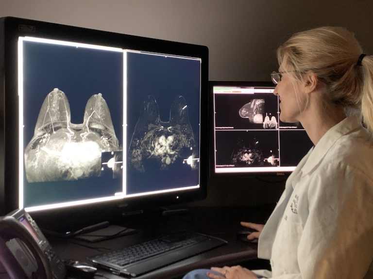
Breast Magnetic Resonance Imaging
Breast Magnetic Resonance Imaging or MRI is a radiation free imaging modality that utilizes a large magnet and radio-waves to generate detailed images of the inside of your breast. Breast MRI has increased diagnostic sensitivity for detecting breast cancers that are not detected mammographically and can help measure the size and extent of a tumor. At Edward Hospital, breast MRI is used for supplemental screening of patients at high risk for developing breast cancer as well as further evaluation of patients already diagnosed with breast cancer. During a breast MRI exam, the patient is positioned face-down on the scanning table and an intravenous injection of contrast dye is administered to the patient while images are being acquired. No compression is necessary however the patient must remain still throughout the exam which takes approximately 30-45 minutes to complete.
Molecular Breast Imaging
MBI or Molecular Breast Imaging is an imaging technique used in women at average risk for breast cancer but who have breasts which are dense on mammography thus making the detection of breast cancer more difficult. MBI, therefore, may be used as a form of supplemental screening. Supplemental screening techniques include MBI, ultrasound and MRI. Each modality has advantages and disadvantages in terms of sensitivity, specificity and the ability to detect cancer without the need for subsequent unnecessary biopsies. At Edward Hospital we have adopted MBI as a supplemental screening modality in average risk patients with dense breasts who wish an additional test to assure the absence of significant abnormalities of the breast while minimizing the risk of unnecessary biopsy due to false positive findings. In patients at high risk for breast cancer due to family history or genetic factors other imaging techniques may be utilized. If you have any questions regarding this procedure, please call the Breast Care Coordinator at (630) 527-3095 (Monday-Friday: 7:30am-4:00pm).
Stereotactic Biopsy
Stereotactic Biopsy is a procedure that utilizes mammography and a computer to sample tissue from a suspicious area of microcalcifications or lesions identified on a mammogram. The computer guides a needle into the area for multiple core samples. The alternative to stereotactic breast biopsy is a surgical removal of the suspicious area through an excisional biopsy. The estimated duration for this procedure is one to two hours. Once the procedure is completed, you would be contacted with results of your biopsy within two to three business days. If you have any questions regarding this procedure, please call the Breast Care Coordinator at (630) 527-3095 (Monday-Friday: 7:30am-4:00pm).
Ultrasound Guided Breast Biopsy
Ultrasound Guided Breast Biopsy is a procedure that utilizes a needle to sample tissue in a lesion that was discovered on your ultrasound. This procedure uses an ultrasound machine to position the needle at the location of the lesion for multiple core samples. The alternative to ultrasound guided breast biopsy is a surgical removal of the lesion through excisional biopsy. The estimated duration for this procedure is one to two hours. Once the procedure is completed, you would be contacted with results of your biopsy within two to three business days. If you have any questions regarding this procedure, please call the Breast Care Coordinator at (630) 527-3095 (Monday-Friday: 7:30am-4:00pm).
MRI Guided Breast Biopsy
MRI Guided Breast Biopsy is a procedure that utilizes a needle to sample tissue in a lesion that was discovered on your breast MRI. MRI stands for Magnetic Resonance Imaging. It uses magnetic fields and radio waves to detect the size and location of breast lesions. This procedure uses the MRI images to position the needle at the location of the lesion for multiple core samples. The estimated duration for this procedure is two to three hours. Once the procedure is completed, you would be contacted with results of your biopsy within two to three business days. If you have any questions regarding this procedure, please call the Breast Care Coordinator at (630) 527-3095 (Monday-Friday: 7:30am-4:00pm).